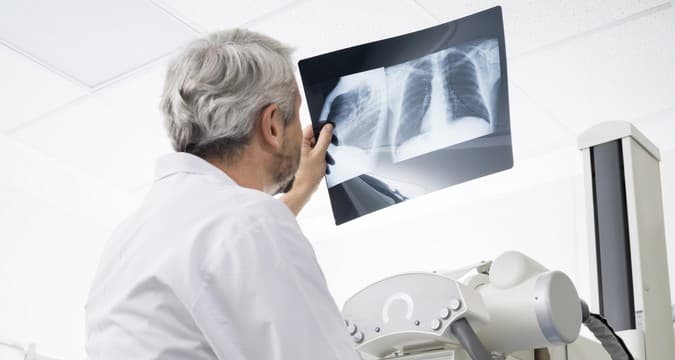
X-ray imaging is a non-invasive diagnostic technique based on the ability of X-rays to penetrate opaque media and structures, be absorbed by them, and project an image onto a special film or screen. With the help of X-ray imaging, almost all bodily systems and organs can be examined, and changes in tissues can be detected even before the onset of pronounced symptoms of a disease. At the GMS Hospital surgical center, X-ray diagnosis is carried out using the most advanced equipment available.

Radiology is a diagnostic method that uses ionized radiation in order to create a localized image of human organs and systems. Our tissues and organs absorb x-rays differently, which shows up in x-ray imagery as varying levels of contrast. This enables doctors to diagnose conditions affecting various organs (lungs, kidneys, etc.) in a short amount of time.
In certain cases, in order to obtain a more precise picture of pathological processes, various contrasting compounds are used. The overall goal is the early detection and diagnosis of organ dysfunctions and illnesses. X-ray technology is used for a wide variety of disorders, and sometimes it is the only effective method of diagnosing serious illnesses.
You can have an X-ray imaging consultation at a convenient time for you, at the GMS Hospital diagnostic center. The main advantages of coming to us for medical care:
You will be given the X-ray images and a copy of the radiologist's report. If necessary, you can immediately arrange for a consultation with a physician specialist.
Thanks to our modern equipment and our doctors' high level of professional training, we can quickly perform the necessary diagnostic examination and obtain sharp, high-quality X-ray images with minimal exposure of the patient to radiation.
At the X-ray and radiology center at GMS we have rooms for CAT, MRT, multi-layer spiral CAT, and X-ray imagery and therapy. The department is equipped with high-tech equipment that allows us to quickly and efficiently perform the whole range of necessary investigations:

GMS Hospital uses high-tech X-ray systems, an orthopantomograph, a photofluorograph, a dental apparatus, and all types of diagnostic studies, including sighting and polyposition, are performed using contrast (barium suspension and iodine-containing agents) and without. Minimal radiation exposure allows us to safely examine patients of all age groups.
The natural contrast in X-ray radiological studies is air and bone and adipose tissue. In some clinical situations, X-ray contrast agents are used to better visualize the organ. Most X-ray procedures at GMS do not require any additional preparation on the part of the patient. However, for the examination of hollow organs (gastrointestinal system), you must first prepare:
Mammography is performed from the 6th to the 12th day of the menstrual cycle. Preparation for hysterosalpingography and for excretory and review urography, as well as for X-rays of the lumbar spine, is similar to that described above. You can get more detailed information about our diagnostic methods and necessary preparatory steps by signing up for a consultation with a doctor, either online or by phone.
Our specialists will contact you